What are the EEG markers of different levels of consciousness? The BIAPT lab is working on EEG-network markers of loss and recovery of consciousness across anesthesia, disorders of consciousness and sleep.
Motifs
Motifs are patterns of inter-connections between nodes of complex networks, with a probability of occurrence that is significantly higher than in randomized networks. As such, functional motifs have been investigated as the basic building blocks of directed networks. The distribution of motifs across a brain network is organized to support information integration and segregation – key properties associated with consciousness. We studied the re-organization of 3-node motifs during loss and recovery of consciousness using nine healthy subjects who underwent a 3 hour anesthetic protocol while 128-channel electroencephalography (EEG) was recorded. We constructed a functional brain network using the weighted and directed phase lag index, on which we calculated the frequency and topology of 3-node motifs. Three motifs were significantly present across participants and levels of consciousness. The topology of long-range chain-like motifs, and short-range loop-like motifs changed significantly across levels of consciousness. Motif topological re-organization preceded (long-range motifs) or accompanied (short-range motifs) the return of consciousness after anesthesia. The full results are published in Scientific Reports: “Brain network motifs are markers of loss and recovery of consciousness”.
We also investigated the prognostic potential of three-node functional network motifs in three patients in a disorder of consciousness, two of whom were exposed to anesthesia. We hypothesized that patient prognosis would be associated with motif topography before, during and after exposure to anesthesia. Next, we compared motif topography to other topographic network properties (network hubs and power topography) and global network properties (graph theoretical network properties and relative power). We predicted that the spatial information provided by topographic network properties would be more predictive of patient outcome compared to global brain network characteristics, which are averaged across nodes in the network. At baseline, the topographic distribution of motifs resembled conscious controls in patients who later recovered consciousness. Strikingly, the reorganization of network motifs, network hubs and power topography under anesthesia followed by their return to a baseline patterns upon recovery from anesthesia, was associated with recovery of consciousness. Our findings suggest that topographic network properties measured at the single-electrode level might provide more prognostic information than global network properties that are averaged across the brain network. The full results are published in Neuroscience of Consciousness: “Brain network motif topography may predict emergence from disorders of consciousness: a case series”.
Types of functional connectivity
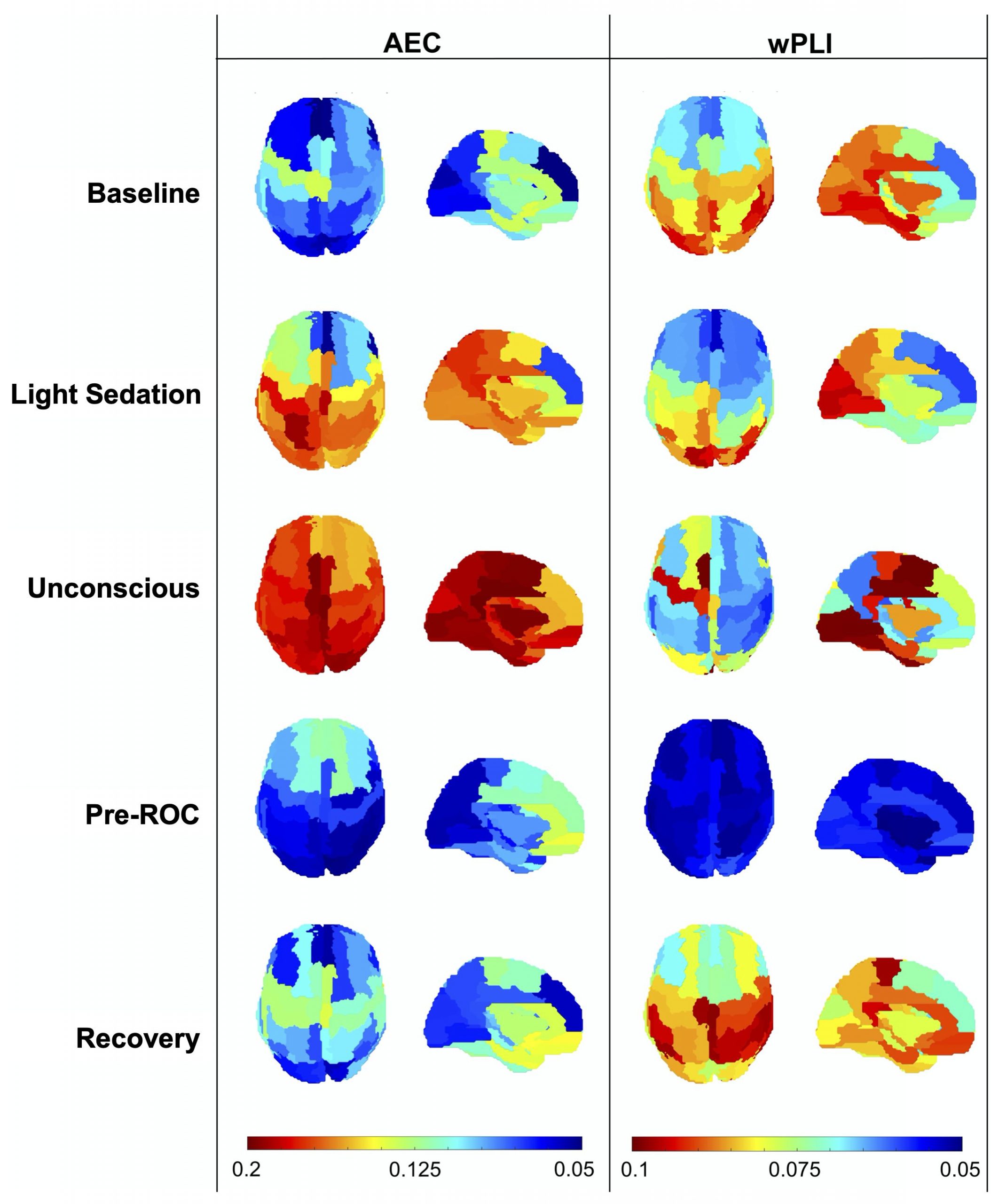
Brain networks can be constructed from both envelope- and phase-based measures of functional connectivity. To date, there have been very few direct comparisons of these techniques using the same dataset. In this project, we compared an envelop-based (i.e. amplitude envelope correlation, AEC) and a phase-based (i.e. weighted phase lag index, wPLI) measure of functional connectivity in their classification of levels of consciousness. We recorded data from nine healthy participants undergoing a three-hour experimental anesthetic protocol with propofol induction and isoflurane maintenance, while 128-channel EEG was recorded. We showed that AEC and wPLI yielded different patterns of global connectivity across states of consciousness, with AEC showing the strongest network connectivity in deep unconsciousness, and wPLI showing the strongest connectivity during full consciousness. Both measures demonstrated differential predictive contributions across participants and used different brain regions for classification. AEC showed higher classification accuracy overall, in distinguishing “deep” from “light” unconsciousness, and when distinguishing between responsiveness and unresponsiveness. Our results show that envelope- and phase-based functional connectivity provide different information about states of consciousness but that (on a group level), AEC is better able to detect relative changes in brain functional connectivity across levels of anesthetic-induced unconsciousness compared to wPLI. The full results were published in NeuroImage: “Differential classification of states of consciousness using envelope- and phase-based functional connectivity”.
Time-resolved functional connectivity
To date, the majority of studies exploring the relationship between functional connectivity and states of consciousness have used a time-averaged approach to generate a representative functional connectivity map of a particular state of consciousness. This traditional time-averaged approach generates representative functional connectivity patterns, but ignores the potentially useful information in the dynamic properties of time-resolved functional connectivity.
The BIAPT lab is currently exploring the functional dynamic architecture of healthy and injured brains and its role in supporting consciousness. Using high-density EEG recorded from healthy controls and patients in a disorder of consciousness, we estimate the strength and directionality of functional connectivity. We characterize the spatiotemporal architecture of functional connectivity and correlate it with the participant’s current level of consciousness, as well as their level of consciousness three months after recording. Our working hypothesis is that individuals who are able to eventually recover consciousness will have a more dynamic architecture of functional connectivity than those that do not.
This project is currently being led by Charlotte Maschke. It is funded by a UNIQUE student fellowship, and the Healthy Brains for Healthy Lives McGill Western Collaboration Grant.

